Dating and Early Pregnancy Scans
This scan is recommended between 6 – 12 weeks of pregnancy and is often recommended if you are unsure of your last menstrual period, have concerns with pain or bleeding, and to determine if you are having more than one baby.
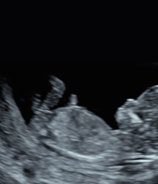
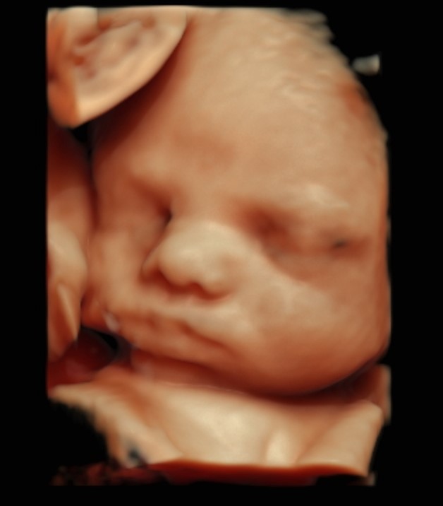
You can expect to see the baby’s heartbeat, however sometimes if the scan is done too early we may not see the heartbeat. This may be perfectly normal, and just too early to detect, so we will bring you back in a few weeks to check the heartbeat and ensure baby is growing as expected.
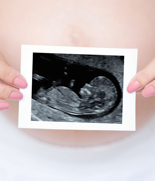
You can also expect to find out the due date of your pregnancy at this scan.
PREPARATION FOR DATING SCAN:
Please drink 2 cups of water an hour before your scan and hold
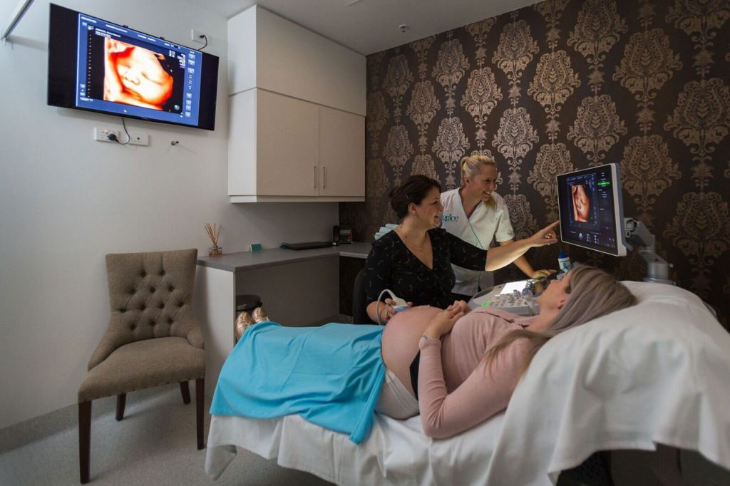
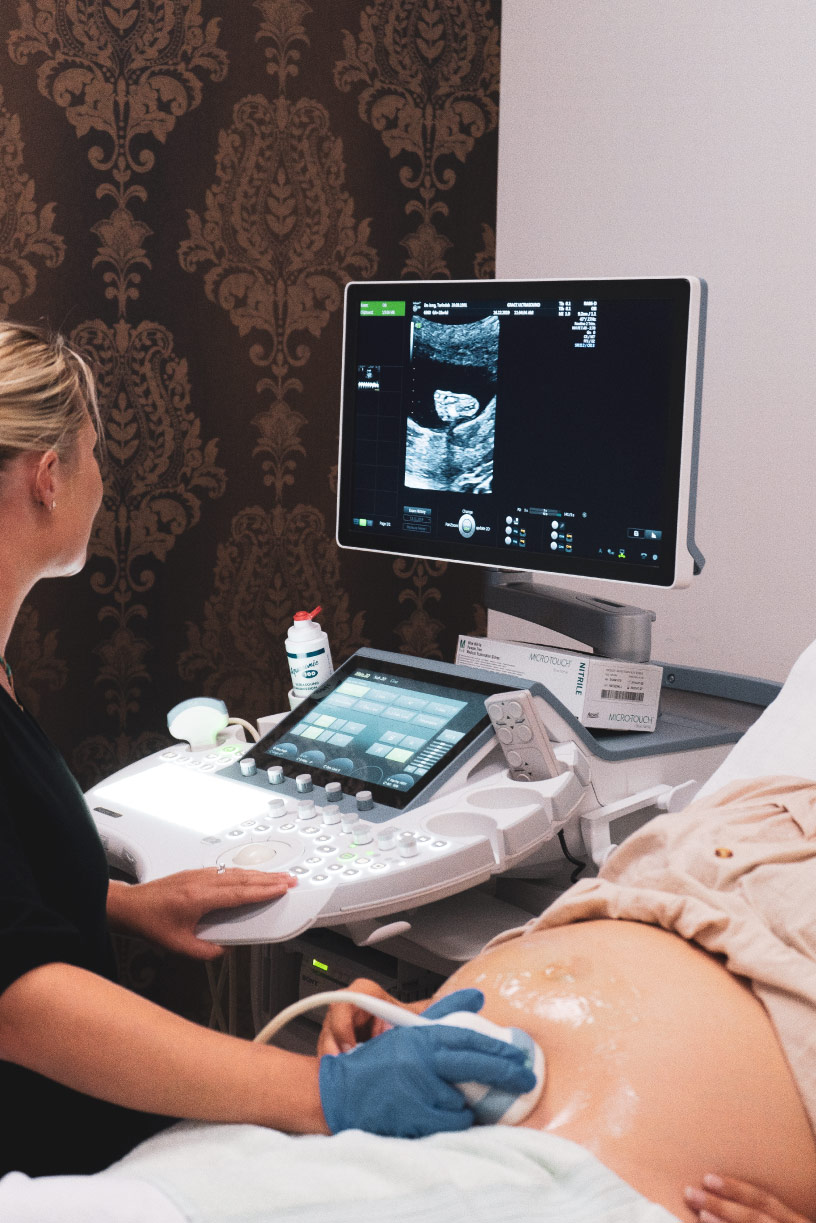
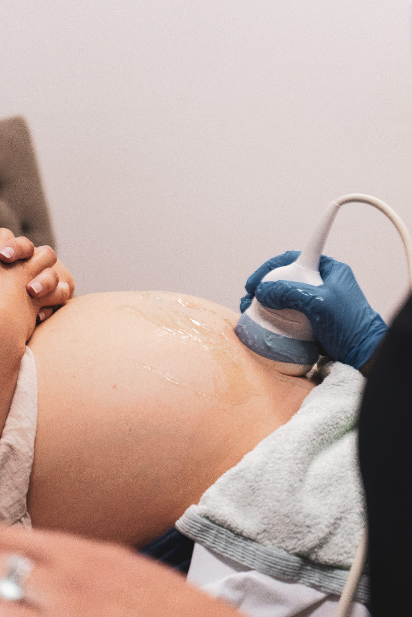
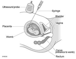
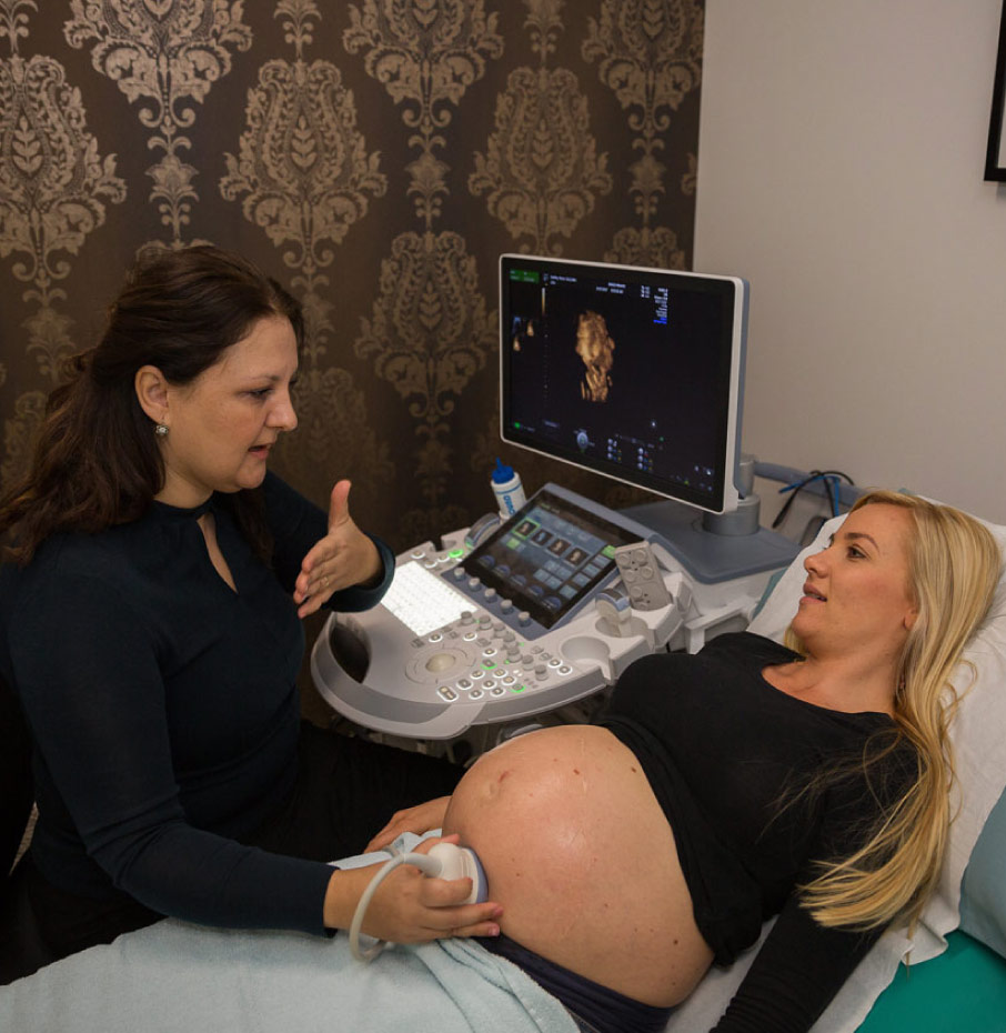
This scan is usually performed abdominally however sometimes we may need to perform a transvaginal scan if your baby is difficult to see or your uterus is tilted backwards. Don’t worry we will always tell you if this is the case and it is our priority to ensure you feel safe and experience minimal discomfort if we need to perform this type of scan. This scan usually takes approximately 10-20 minutes